Yorgia
|
Yorgia Temporal range: Ediacaran |
|
|---|---|
 |
|
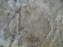
| Fossil of Yorgia waggoneri | |
| Scientific classification | |
| Kingdom: | Animalia |
| Phylum: | Proarticulata |
| Family: | Yorgiidae |
| Genus: | Yorgia |
| Species: |
Y. waggoneri Ivantsov, 1999 |
Yorgia waggoneri is a discoid Ediacaran, resembling a transition between the organisms Dickinsonia and Spriggina. It has a low, segmented body consisting of a short wide "head", no appendages, and a long body region, reaching a maximum length of 25 cm (9.8 in). It is classified within the extinct animal phylum Proarticulata.
The generic name Yorgia comes from the Yorga river on the Zimnii Bereg (Winter Coast) of the White Sea, where the first specimens were found. The specific name in honor of U. S. paleontologist Ben Waggoner, who found the first specimen.
The body plan of the Yorgia and other proarticulates is unusual for solitary (non-colonial) metazoans. These bilateral organisms have a segmented metameric body, but left and right transverse elements (isomers) are organized in an alternating pattern relatively to the axis of the body – they are not direct mirror images. This phenomenon is described as the symmetry of gliding reflection, which is a characteristic also found in the similar Spriggina. Some proarticulates (Yorgia, Archaeaspinus) demonstrate obvious asymmetry of left and right parts of the body. Yorgia’s initial right isomer is the only one which spreads far towards the left side of the body. This lack of true bilateral symmetry, along with other considerations, has led some scientists to suspect that the organism falls in a sister group to the eumetazoa (i.e. all animals except Parazoa).
Imprints of the Yorgia waggoneri has been found in the rocks of Vendian period (Ediacaran) White sea region of Russia, dated around 555.5 Ma. and Yorgia sp. has been found in the Central Urals of Russia and Flinders Ranges, Australia. Most body imprints of Yorgia have in the past been primarily preserved on the sole of sandstone beds in negative relief. Other Yorgia fossils show internal structure in the original organism, showing two symmetrical rows of nodules, a central tube, rib like tubes, and a semicircular shape with a hole in the circle centre positioned towards the head end. This structure has been interpreted as the impression of gonads, intestine and mouth.
...
Wikipedia
